UL
Written by: Uta Leyke-Hess
Expert review by: Rupa Dainer
Published: 13. April 2024
Expert review by: Rupa Dainer
Published: 13. April 2024
Reading time ca.
Getting regular breast cancer screenings improve the chances of early detection. This is important because finding breast cancer as early as possible enables prompt and effective intervention. Not sure what breast cancer screenings involve? Well, you’ve probably heard of women getting mammograms. A mammogram is the image of a breast produced during a mammography screening. To confirm a breast cancer diagnosis, your healthcare provider may follow up a mammography screening with other kinds of imaging techniques.
In this article, you’ll learn about:
- techniques doctors use to confirm a breast cancer diagnosis
- why mammograms are preferred for breast cancer screening
- other screening techniques like ultrasound, magnetic resonance imaging (MRI), molecular breast imaging (MBI), and the use of biochemical biomarkers.
How is breast cancer diagnosed?
A breast cancer diagnosis can be made in multiple ways. The most common approach is the use of imaging techniques. The preferred method to screen for breast cancer is mammography. If your mammogram shows any signs that could point to cancer, then your radiologist may recommend other imaging techniques. These include ultrasound, magnetic resonance imaging (MRI), or molecular breast imaging (MBI).
In addition to these imaging techniques, a breast cancer diagnosis can be made by looking for biochemical biomarkers. These biomarkers are produced by cancer cells, or by healthy cells in response to cancer. Detecting these biomarkers may indicate the presence of cancer in the body.
Biomarkers can provide useful information about cancer which can be used both in breast cancer diagnosis as well as breast cancer treatment. These biomarkers can be obtained by taking a sample during a biopsy, or by having bloodwork done.
What is a mammography?
A mammogram is the most commonly used tool to screen for breast cancer. To obtain a mammogram, first the breast is placed on top of a flat support plate and is gently compressed with another plate. Next, an X-ray machine takes an image of the breast from multiple angles.
This results in a mammogram, which can be analyzed by a radiologist. The radiologist searches for areas that look different from normal tissue. They will assess the size, shape, and contrast of the abnormal area/s to determine if it is harmless or potentially cancerous.
Your radiologist may also order additional imaging to confirm or further investigate the results.
What types of mammograms are used to diagnose breast cancer?
There are various types of mammograms. 3D mammography is rapidly proving to be more sensitive that 2D mammography and is now routinely covered by most commercial insurance. The possible types of mammograms are:
- Conventional mammogram (2D): this technique captures the x-ray image on a film. It shows tissues such as fat in shades of gray and tissues such as glands or tumors in a shade of white.
- Digital mammogram: this technique is similar to a conventional mammogram. The difference is that the X-rays get converted into electronic signals which are passed along to a computer. The computer converts these signals into digital images. This technique has the advantage that the image can be manipulated to have a better view of possible abnormalities.
- 3D mammography, also known as digital breast tomosynthesis: this technique creates a 3D representation of the breast. It does this by producing X-rays at different angles to the breast.
What is the alternative to a mammogram?
Although mammograms are often used for breast cancer screening, there are plenty of other imaging techniques that can provide more information on whether or not cancerous cells are present. These techniques are mostly done in addition to a mammogram to help confirm a breast cancer diagnosis. Some techniques include:
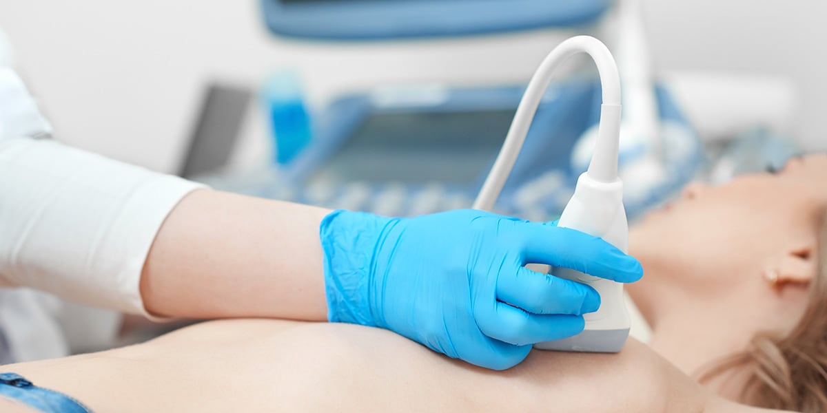
- Ultrasound: this technique uses sound waves to create images of the breast. It is usually done after a mammography and can help confirm if unusual areas noted on the breast x-ray are solid or fluid-filled. Solid areas are more likely to be masses, while fluid filled areas may be cysts or signs of infection. Early-stage breast cancer on ultrasound can appear as a dark spot with a slightly lighter color and an irregular shape.
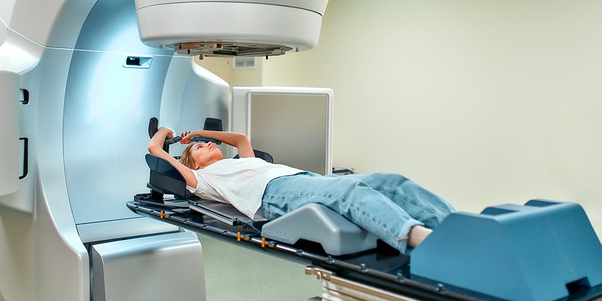
- MRI: this technique provides breast cancer images using magnets and radio waves. Breast MRIs are often used to screen women who have a higher risk of developing breast cancer.
- Biopsy: with this technique, cells are extracted from the breast or the lymph nodes in the armpit. The extracted cells are then sent to a lab to see if they are cancerous. A biopsy is often performed with the help of an ultrasound.
- Molecular breast imaging (MBI): for this technique, a small amount of radioactive tracer needs to be inserted in the breast. With the help of a special camera, your radiologist can distinguish between healthy tissue and abnormalities. This diagnostic tool is often used for women with more dense breast tissue and women who have a higher risk of developing breast cancer or a recurrence of breast cancer.
- Diagnostic biomarkers: with this technology, certain molecules can be detected that indicate that a cancer is present. This can be done by examining a tumor biopsy or the blood.
- Position emission tomography (PET) scan: this technique requires the injection of a small amount of radioactive glucose, after which a scanner can make computerized pictures of the body. As cancer cells often take up more glucose than normal cells, the pictures can point to cancerous processes in the breast.
- Computerized tomography (CT) scan: this procedure creates pictures from different angles to create a 3D view of the tissue using X-rays. A dye might be used to make specific tissues more visible in the image.
How is the stage of breast cancer determined?
Breast cancer can be divided into different stages in order to indicate the severity of the disease. The stage of the cancer can influence the treatment and outlook of the disease. For breast cancer, there are five general stages:
- Stage 0: This means there are cancer cells present but they have not spread outside the ducts or lobules of the breast.
- Stage 1: This means that the tumor is now considered invasive, and has spread to normal breast tissue. It is only present in the breast and hasn’t spread to the lymph nodes, or it has only spread to a nearby lymph node without being present in the breast at all.
- Stage 2: This means that the cancer may have spread to a few lymph nodes located nearby, or has grown larger than 5 cm without spreading to nearby lymph nodes
- Stage 3: This means that the tumor is slightly larger than in stage 2 and that it has spread to several lymph nodes or other tissues nearby such as the chest wall.
- Stage 4: This is the most advanced stage of breast cancer. It means that the cancer has spread to different parts of the body. The survival rate of people with stage 4 breast cancer is significantly lower than that of other stages.
What does inflammatory breast cancer diagnosis mean?
Inflammatory breast cancer is a rare condition that often progresses rapidly. This type of breast cancer blocks the lymph vessels in the skin of the breast, which causes the breast to look swollen and red. It is often diagnosed at a younger age, unlike other forms of breast cancer in which the average age of diagnosis is usually around 62.
Inflammatory breast cancer is usually diagnosed after a mammogram and an ultrasound of the breast and the lymph nodes. A PET scan, CT scan, or bone scan may be performed to see whether or not the cancer has spread.
In summary
The diagnosis of breast cancer can be made using various techniques. The preferred way of screening for breast cancer remains the mammogram, although this technique is often followed by other imaging techniques that can be used to confirm breast cancer such as an ultrasound, MRI, or MBI. Which technique is used after a mammogram depends on the individual’s situation.
FAQ
References
UL
Written by Uta Leyke-Hess
Medical writer
Uta Leyke-Hess holds a degree in business administration with a strong focus on healthcare. Since 2015, she has been supporting various medical websites by creating high-quality, yet easy-to-understand content with the help of a team of qualified external writers she has assembled.